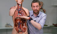
Anatomy
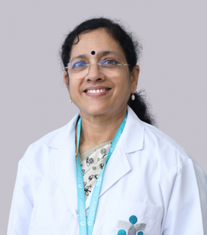
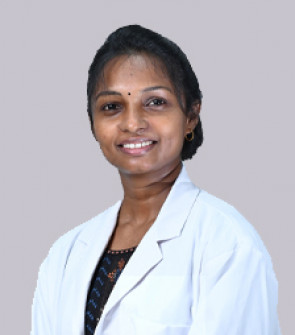
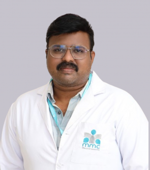
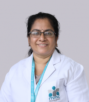
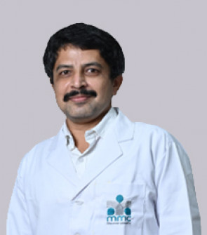
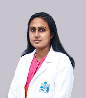
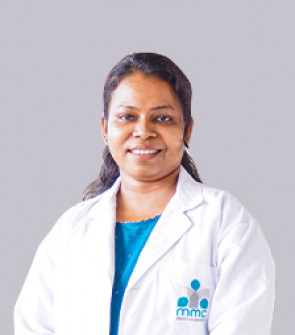
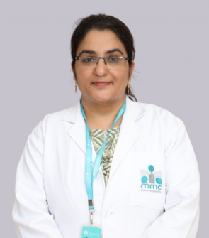
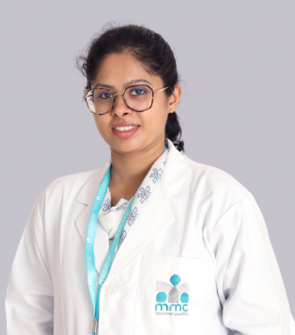
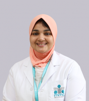
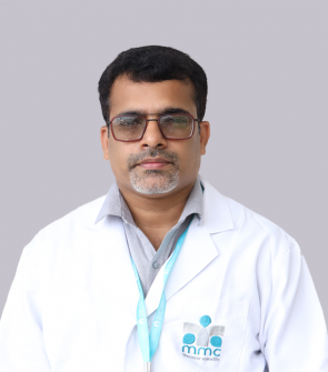
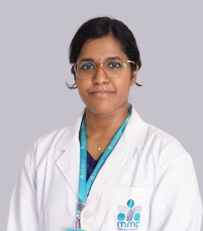
Doctors

About the Department
- Malabar Medical College, Department of Anatomy was established in 2009
- Primarily a teaching department and responsible for teaching anatomy and guiding 150 Medical, 100 Dental andmany BSc Nursing and paramedical students.
- The Department try to inculcate a love for the subject by using different modalities of teaching and interactive sessions and make their tenure in the department informative, enjoyable and a memorable one
- Anatomy taught in three ways : Gross anatomy by cadaver dissection, Microanatomy by projection of histology slides and Embryology using charts and models
Departmental Focus
Academics
- To provide the basic knowledge of Anatomy for building a strong foundation for a clinical career.
- To imbibe professionalism into the budding doctors.
- To instill a research oriented approach at the undergraduate level
- To improve continually the efficiency of learning for our students
- To strive for excellence in the Anatomy Teaching Programmes
Infrastructure
Dissection Lab – Department consists of an excellent, spacious and well ventilated dissection hall with an attached cadaver storage and specimen handling room. Seperate locker rooms are provided for girls and boys to keep their belongings. LCD projector with big screen is being provided in the dissection hall for giving a preview on dissection.
A well equipped Histology lab with around 90 microscopes and good quality sections of all tissues. Two preparation rooms with all the equipments required for preparation of slides and its staining
A well spacious Lecture hall with LCD projector
2 Demo rooms with LCD projectors
Museum with about 250 specimens including soft tissues, bones & models
Department library with good collection of reference books
Publications
RESEARCH PUBLICATIONS BY FACULTIES OF THE DEPARTMENT
Dr. Geetha. K. N
1. Mehta UK, Geetha KN* , Gaikward M, Chavan L _ Posterior arch anomalies of Atlas and its clinical significance International Journal of Medical Research & Review- 2014, vol.2:361-366.
2. Dashrath Haribhau Pimple, Geetha K N*, Karuna N Katti, G V Kesari – Morphological Study of External Ear in Mentally Retarded & Healthy Subjects-Journal of Medical & Health Sciences 2013,vol.2(4):92-97.
3. Geetha K N*, Saroj V Kotwaliwale – Effectiveness of Fluorescent in Situ Hybridization (Fish) over conventional Karyotyping as a diagnostic tool for pre natal detection of aneuplody of chromosome 21 – The Journal of Anatomy 2014, photon 114: 149-153.
4. Geetha K N*, Patel S .C, Chavan L.N, Charushila Shinde- Hand digit ratio (2D:4D) & Sexual dimorphism in different age groups- Journal of Clinical Research Letters – 2012 Vol 3 (1):16-18.
5. Chavan L.N, Geetha K N*, Karuna Katti, Shinde C.D – Estimation of Stature from Foot Dimensions of School age group Children in Maharashtra State- International Journal of Medical & Clinical Research -2012 vol 3 (2) : 121- 126.
6. Lalita N Chavan. Geetha K N* , Nilesh Nangri, Roshan S, Vitthal Karkara, Rajesh Dwivedi – Estimation of Stature , Age & Sex from Foot Dimensions in 18-20 yrs of age of students in Maharashtra State ,India- International Journal of Medical Research & Review- 2014, vol.3 (3) : 71-76.
7. Ashish Kulkarni, Geetha K N* – Variation in branching pattern in Facial artery: An anatomical study in 50 adult cadavers. The Journal of Anatomy- 2013 Photon 113 : 132-134.
Dr. B S Rathna
1. Gopalakrishna.K1 , B.S.Rathna* , The study on the incidence and direction of nutrient foramina in the diaphysis of femur bone of south Indian origin and their clinical importance. IJBLS- International journal of basic and life sciences. 2014, Vol- 2 Issue-2, April,11-19.
2. Gopalakrishna. K 1, Deepalaxmi S 2, Somashekara S.C 3, Rathna B S* An anatomical study on the position of Mandibular Foramen in 100 dry mandibles. International Journal of Anatomy and Research, Int J Anat Res 2016, Vol 4(1): 1967-71. ISSN 2321-4287. DOI: http://dx.doi.org/10.16965 / ijar.2016.122.
3. Gopalakrishna K, Deepalaxmi S, Somashekara S C, Rathna B S*- A cadaveric study on morphological variations of fissures and lobes in the human lungs and its clinical significance. Journal of Experimental and Clinical Anatomy ‐.Volume 16, Issue 1, January-June 2017; 16:7-11.
Dr. K Meera
1. Kugananthan.M*, Sadeesh.T, D’Silva. H, Anbalayan.J, Sudha Rao- Histological study on the Obliteration process of Ductus Arteriosus in still born fetuses. Jr Dental and Medical Sciences.2014:13(7);28-31.
2. Rao.S, Kugananthan.M *, K.Sujatha. H.R. Krishna Rao- A study on branching pattern of arch of aorta with its embryological significance and review of literature. Int.Jr.of Anatomy and Research.2017,5(1):3516-20.
3. N.Shakunthala, Kugananthan.M *, K.Sujatha, H.R.Krishna Rao- A radiological anatomical study on Cadaveric Kidneys to trace the course of polar arteries to Kidneys. Int.Jr.Anat Res 2016, 4(4): 3156-60
4. Kugananthan.M *, Anbalayan.J, Sudha Rao, Hydrina D’Silva, Sadeesh.T- Congenital diaphragmatic hernia. A case report. JMed Sci.2014:(3(2), 80 - 83
5. VedhaShanmugaSundaram, Kugananthan.M *, Sadeesh.T, Rijied Thompson Swer, Aruchandra Singh- A rare variant of bilateral brachial plexus. A case report. Jr.of Int Academic Res for multi disciplinary-2014 :2(7);546-50.
6. Prabhavathy.G, Sadeesh.T, Kugananthan.M *, Chandra Philip X, Anbalayan.J. Histomorphological study of Hyrtt’s anastomosis in pregnancy induced hypertension and small for gestational age- A clinical correlation. Biomedicine-2020;40(2) : 123-127
Dr. Srikrishna Ganesh Kulkarni
1. “Dermatoglyphics in Primary Hypertensive patients” International Journal of Pharma & Bio Sciences. 2014 Jan, 5(1): (B) 53-58.
2. An Anatomical study on the Frequency of Nutrient Foramina in the Shaft of Tibia Bone from Indian population with its clinical importance. International Journal of Innov Research and studies. 2015, July, Vol.4, Issue 7,16-26.
3. “Study on variability in the location of nutrient foramen in fibular diaphysis of Indian population” Scholars Journal of Applied Medical Science, August 2015;3(5D):2091- 2095.
4. “The Evaluation of Variation in the Length of Styloid Process of Indian Population and its applied importance”. International Journal of Allied Medical Science and Clinical Research . Vol- 3 (3)2015 (373-378)
Dr. Gopalakrishna. K
1. Gopalakrishna K*,KashinathaShenoy M, Preetha., ‘Meningo-Orbital Foramen-In South Indian Dry Skulls and Its Incidence’. RJPBCS, Vol-5(1) Jan-Feb 2014, Page 729-734.
2. Gopalakrishna.K1*, B.S.Rathna2 , The study on the incidence and direction of nutrient foramina in the diaphysis of femur bone of south Indian origin and their clinical importance. IJBLS- International journal of basic and life sciences. 2014, Vol- 2 Issue-2, April,11-19.
3. KashinathaShenoy*, Gopalakrishna.K., A clinical profile of fungal corneal ulcer in tertiary eye care hospital in coastal Kerala. International journal of medical and applied sciences. 2015, Volume 4, Issue 1, page 25-29.
4. DeepalaxmiSalmani*1, Suja Purushothaman2,*, Gopalakrishna3, Laveesh Ravindran4, Santhi ReghuNath5, BisharPushkar6., A study of Dermatoglyphics in relation with blood groups among first year MBBS students in Malabar Medical College., Indian Journal of Clinical Anatomy and Physiology, July-September 2016;3(3);348-350. DOI: 10.5958/2394- 2126.2016.00079.7
5. Gopalakrishna. K *1, Deepalaxmi S 2, Somashekara S.C 3, Rathna B.S 4. An anatomical study on the position of Mandibular Foramen in 100 dry mandibles. International Journal of Anatomy and Research, Int J Anat Res 2016, Vol 4(1): 1967-71. ISSN 2321-4287. DOI: http://dx.doi.org/10.16965 / ijar.2016.122.
6. Gopalakrishna K*, Deepalaxmi S, Somashekara SC, Rathna BS. A cadaveric study on morphological variations of fissures and lobes in the human lungs and its clinical significance. Journal of Experimental and Clinical Anatomy ‐.Volume 16, Issue 1, January-June 2017; 16:7-11.
Mrs. Sreekala M A
1. Gopalakrishna K , Sreekala M A*, B S Rathna- A study on the incidence and direction of nutrient in South Indian humeral diaphysis and their clinical importance/ Research and Reviews: Journal of Medical & Health Sciences- 2014: vol 3(1):71-76.
2. Gopalakrishna K, Sreekala M A*, B S Rathna -The study on the incidence and direction of nutrient foramina in the radius bone of South Indian Ulnar diaphysis and their clinical importance -International Journal of health sciences and Research ,2014.Vol-4, (3):30-37.
Dr. AarabhyJayan
1. JAYAN, A., JS, K. and GOPI, J., 2021. Accessory Head of The Flexor Pollicis Longus: A Cadaveric Study on The Gantzer Muscle. Journal of Clinical and Diagnostic Research.
Dr. Soumya. R
1. Ramakrishnan S, Kunjunni KT, Varghese S. A comparative study on segmental micro-anatomy of the human fallopian tube.Natl J Clin Anat 2021;10:46- 50
Other Activities
Involved in teaching of MBBS, BDS, BScNursing, Bsc Optometry, BSc MLT,BPT,DRTcourses
Teaching Learning Methods have been updated in accordance with the new Competency Based Medical Education.
Seminar presentations for students are done regularly after which an interactive session is conducted between faculties & students for clarifying the doubts & understanding concepts
Small group discussions are being conducted frequently from dissection hall and active participation of each & every student is ensured
Clinical Oriented questions are given related to each topic and doubts regarding it are clarified
Vertical integration for clinically relevant topics are conducted monthly where clinicians from various specialities give interesting talks on the subject including the videos of various clinical cases & procedures .Students were also taken to wards to observe the Bedside clinical evaluation by clinicians and understand the rapport between doctor & patient.
Periodic feedback are collected from table representatives from each dissection table where they can address the difficulties faced by them and appropriate rectifying measures are taken accordingly.
SLOs & relevant diagrams for each topic is given.
After each internal exams students are categorized according marks.Intensive training , great attention& care are given for weak performers
Table wise mentoring is done regularly by table teachers where mentees are being monitored continuously.Students are free to communicate academic, personal, financial, social or any problems with the mentors which will be held highly confidential and solutions are sorted out .Feedback of each mentee is given to parents periodically. Parents are also free to contact mentors if they have any queries.
Model making contest for embryology topics was conducted to make the students study the topics in a more interesting& simplified way Students were divided in to groups and each group was asked to select a particular topic.They were given 1 hour time to make the model. Representatives from each group was asked to present the topic.All the groups came out with wonderful models which helped them a lot in stretching their imaginations and clarifying the doubts.


